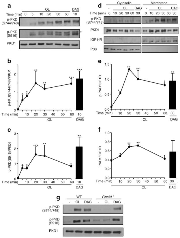
Fig. 4.
Oleate phosphorylates PKD1 in a GPR40-dependent manner. (a) Representative immunoblots of phospho-Ser-744/748 (S744/748) PKD, phospho-Ser-916 (S916) PKD and total PKD1 in protein extracts of INS832/13 cells exposed at indicated time to 16.7 mmol/l glucose ±0.5 mmol/l oleate or 0.1 mmol/l 1,2-dioctanoyl-DAG. The arrow shows S916 PKD based on the expected molecular mass. (b, c) Quantitative measurements of phospho-Ser-744/748 PKD (b) and phosho-Ser-916 PKD (c) in INS832/13 cells. (d) Representative immunoblots of phospho-S744/748 PKD and total PKD1 in cytosolic and membrane fractions of INS832/13 cells exposed to 16.7 mmol/l glucose ±0.5 mmol/l oleate (OL) or 0.1 mmol/l 1,2-dioctanoyl-DAG. Immunoblots of P38 and IGF1R were used as cytosolic and membrane markers, respectively. (e, f) Quantitative measurements of phospho-S744/748 PKD (e) and total PKD1 (f) in cytosolic fractions. (g) Representative immunoblots of S744/748 PKD, S916 PKD and total PKD1 in WT and Gpr40−/− islets exposed for 30 min to 16.7 mmol/l glucose ±0.5 mmol/l oleate or 0.1 mmol/l 1,2-dioctanoyl-DAG. Data are mean±SEM of three independent experiments. *p<0.05, **p<0.01, and ***p<0.001 compared with time 0

