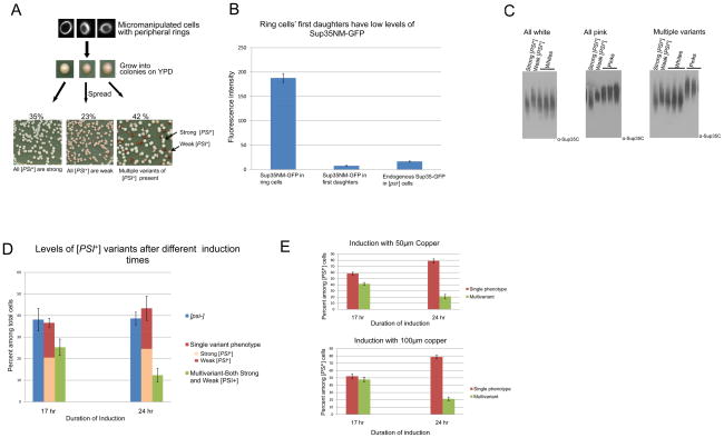
Figure 2. More than one [PSI+] variant can arise from a single cell following de novo induction of [PSI+].
A. One ring cell can give rise to progeny that are all strong [PSI+], all weak [PSI+] or a mixture of strong and weak [PSI+]. [PSI+] was induced de novo by over expressing Sup35NM-GFP in [PIN+] [psi−] cells. Cells with rings were micromanipulated and grown on YPD plates for 3 days where Sup35NM-GFP expression was turned off. The resulting colonies were suspended in water and spread on YPD. The types of [PSI+] variants present in the ring cell progeny were determined from the color of these colonies (>500 ring containing cells were micromanipulated). Only ring cells giving rise to some [PSI+] progeny are depicted. B. Ring cells’ first daughters have low levels of Sup35NM-GFP. Comparison of endogenous Sup35-GFP fluorescence in [psi−] cells with cytoplasmic Sup35NM-GFP fluorescence in ring containing mother cells and their first daughters. Images of GF-658 (MAT α ade1–14 ura3–52 leu2–3,112 trp1–289 his3–200 SUP35-GFP) were taken for the [psi−] cells. Ring cells and daughters of ring cells were imaged from L1749 (MAT a ade1–14 ura3–52 leu2–3,112 trp1–289 his3–200) transformed with pCup-SUP35NM-GFP after induction until ring stage. After induction and the appearance of rings, cells were washed with water and grown in YPD for ~ 3 hrs. Images were then acquired and quantified from three different trials of 10 cells each (see Experimental Procedures). All cells were imaged under identical conditions Bar graphs represent the average fluorescence intensity of Sup35-GFP and Sup35NM-GFP in the respective cells. The level of Sup35-GFP was reduced about 25% relative to the level of untagged Sup35 in Western blots (data not shown). This may reflect differential degradation in the lysate. C. Confirmation that different color colonies have variants with different sized Sup35 oligomers. Cell lysates were prepared from colonies shown at the bottom of 2A. Crude lysates were treated with 2% SDS at room temperature and SDS resistant oligomers were analyzed by SDD-AGE analysis. Lysates from strong [PSI+] (L1762) and weak [PSI+] (L1758) variants were run as controls. Left panel shows results from ring cells whose [PSI+] progeny were all white, center shows results from ring cells whose [PSI+] progeny were all pink and right panel shows results from ring cells whose [PSI+] progeny were both white and pink. D. [PSI+] variant establishment varies with the duration of Sup35NM protein induction. [PIN+] [psi−] cells containing the Sup35NM-GFP plasmid were grown in 50 μM CuSO4 at 30°C for 17 or 24 hrs to induce [PSI+]. Ring aggregate containing cells were then isolated and progeny examined. Ring cells gave rise to [PSI+] variants with a single phenotype more frequently after 24 vs. 17 hrs of Sup35NM-GFP expression. Each bar represents standard error of more than 3 trials of at least 100 viable ring cells. Data includes all ring cells whether or not they gave rise to any [PSI+]. E. Increasing expression of Sup35NM-GFP does not alter the variant establishment. Individual ring cells were micromanipulated after 17 or 24 hrs of induction with 50 μM or 100 μM of CuSO4 and the [PSI+] variant types arising from these cells were determined. No significant difference was observed in the relative frequencies of [PSI+] variants whether 50 or 100 μM CuSO4 was used. Each bar indicates standard error of more than three trials of 100 viable ring cells. Data shown includes only ring cells giving rise to some [PSI+] progeny. (For all experiments starting OD at 0 hrs=0.1, OD at 17 hrs~1.0 and OD at 24 hrs ~2.0 unless otherwise indicated).

