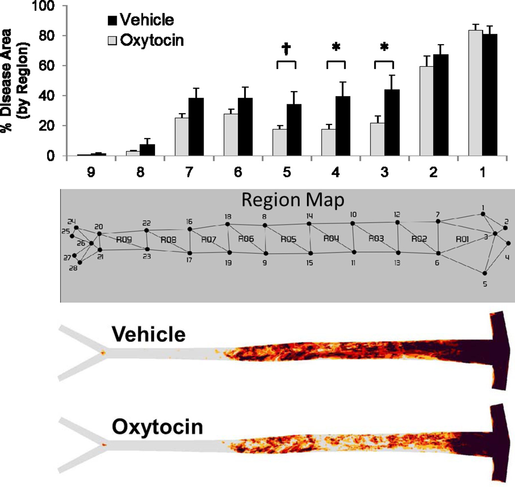
Figure 2.
Aortic atherosclerosis lesion prevalence map and bar graph showing quantitative lesional area within assigned aortic regions in WHHL rabbits chronically infused with OT or saline for 16 weeks. There was a significant decrease in atherosclerosis in the thoracic aorta identified as regions 3, 4 and 5 and trends toward lower extent of disease in the upper abdominal aorta (regions 6 and 7). No differences were observed in the aortic arch (regions 1 and 2). *p ≤ 0.05 between Groups, † p = 0.088.

