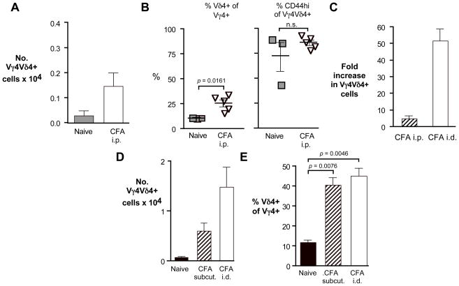
Fig. 4. The route of immunization influences the strength of the response of the Vγ4Vδ4+ subset to CFA.
A–C. C57BL/6 mice were either uninfected (naïve controls), or given an intraperitoneal (i.p.) injection of CFA emulsified in PBS on days 0 and 21. Cells were stained for flow cytometric analysis 26 days after the first injection. A. Peritoneal Vγ4Vδ4+ cell numbers obtained. B. Percent of peritoneal Vγ4+ cells co-expressing Vδ4 (left) and percent of peritoneal CD44-high Vγ4Vδ4+ cells (right). C. The fold-increase in intraperitoneally (i.p) or intradermally (i.d.) immunized mice was calculated by dividing the number of Vγ4Vδ4+ cells obtained in each CFA stimulated mouse by the average obtained per mouse in naïve controls. D–E. C57BL/6 mice were injected twice as for intradermal CFA immunization, but subcutaneously at the scruff of the neck, and cells from the axillary and inguinal lymph nodes were combined and analyzed on day 26. Lymph nodes were pooled for the naïve controls, whereas subcutaneously or intradermally injected mice show the average obtained from 4–5 animals. D. Vγ4Vδ4+ cell numbers obtained from lymph nodes of mice immunized by subcutaneous injection of CFA. E. The percent of Vγ4+ cells co-expressing Vδ4 from the same samples shown in D.

