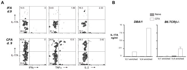
Fig. 8. Vγ4Vδ4+ cells induced to express IL-17 do not co-express IFNγ or IL-2, although some co-express TNFα.
A. C57BL/6 mice were immunized by intradermal injection of CFA or IFA, and lymph nodes harvested on day 9. The panels show a typical example of a flow cytometry panel from an individual mouse. B. Draining lymph nodes were taken on day 20 from DBA/1LacJ or B6.TCRβ−/− mice that were either left untreated, or that received intradermal CFA injections on days 0 and 14. Cells from 3 mice were pooled, passed over nylon wool to enrich for T cells, and negatively selected using MACS beads to enrich for either Vγ1+ or Vγ4+ cells. Cells were cultured on wells coated with anti-Cδ monoclonal antibody, and the presence of various cytokines measured using a multi-plex cytokine bead assay with known standards. Cells from DBA/1 mice show the results from single cultures; error bars for cells from B6.TCRβ−/− mice show the range of values obtained from duplicate wells tested separately.

