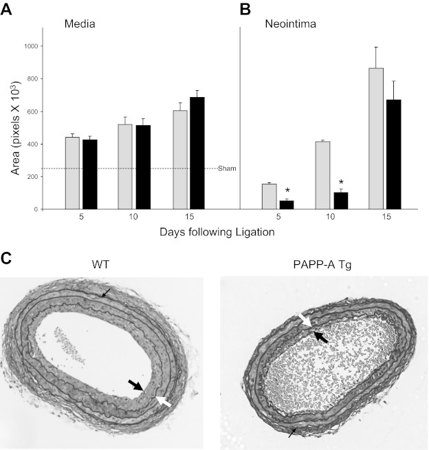
Fig. 2.
Medial (A) and neointimal (B) area in wild-type (WT; gray bars) and PAPP-A Tg mice (black bars) following unilateral carotid ligation. Results are means ± SE of 8–12 mice. Dashed line indicates sham values in medial area. *Significant difference between WT and PAPP-A Tg mice at P < 0.0001. C: sections of carotids 10 days following unilateral ligation stained with Verhoff von Giessen. Thick black arrows indicate the neointima, thick white arrows indicate the internal elastic lamina (IEL), and thin black arrows indicate the external elastic lamina (EEL). Neointimal area was defined by IEL minus luminal area. Medial area was defined by EEL minus IEL.

