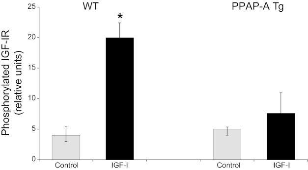
Fig. 6.
IGF-I-stimulated IGF-I receptor (IGF-IR) phosphorylation ex vivo. Two days postligation, carotids from WT and PAPP-A Tg mice were harvested and then treated without (gray bars) or with 50 nM IGF-I (black bars) for 10 min. Western immunoblotting for phosphorylated IGF-IR was performed and analyzed as described in materials and methods. Results are means ± SE (n = 5 mice). *Significant IGF-I effect at P < 0.05.

