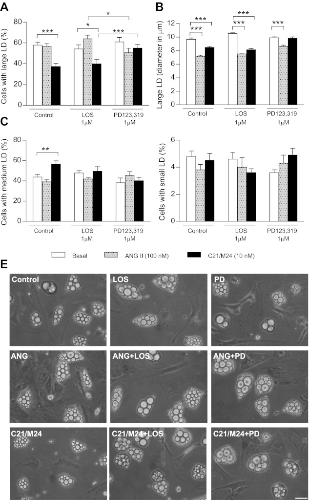
Fig. 4.
Effect of selective ligands of type 1 and type 2 receptors on morphology of adipocytes from SC rat adipose tissue in primary culture. Preadipocytes were plated in 35-mm Petri dishes at a density of 1.5 × 10⁴/cm2 and cultured until day 13 as described in research design and methods in the absence (control) or presence of ANG II (100 nM) or C21/M24 (10 nM) alone or in combination with losartan (1 μM) or PD123,319 (1 μM). A and B: histograms illustrating percentage of cell types exhibiting large LD (A) and their mean diameter (B). C and D: histograms illustrating percentage of cells types exhibiting medium LD (C) and small LD (D). Values were obtained from data analyses (histograms and fitting curves) constructed as described in research design and methods and in Supplemental Data. Data are means ± SE of LD from 54 images taken from 3 different Petri dishes from 3 different experiments. Statistical analyses were performed using two-way ANOVA followed by Bonferroni multiple comparisons. Statistical significance vs. respective control: *P < 0.05, **P < 0.01, and ***P < 0.001. E: adipocytes cultured for 13 days in the absence (control) or presence of ANG II (100 nM) or C21/M24 (10 nM) alone or in combination with losartan (1 μM) or PD123,319 (1 μM). Scale bars, 40 μm.

