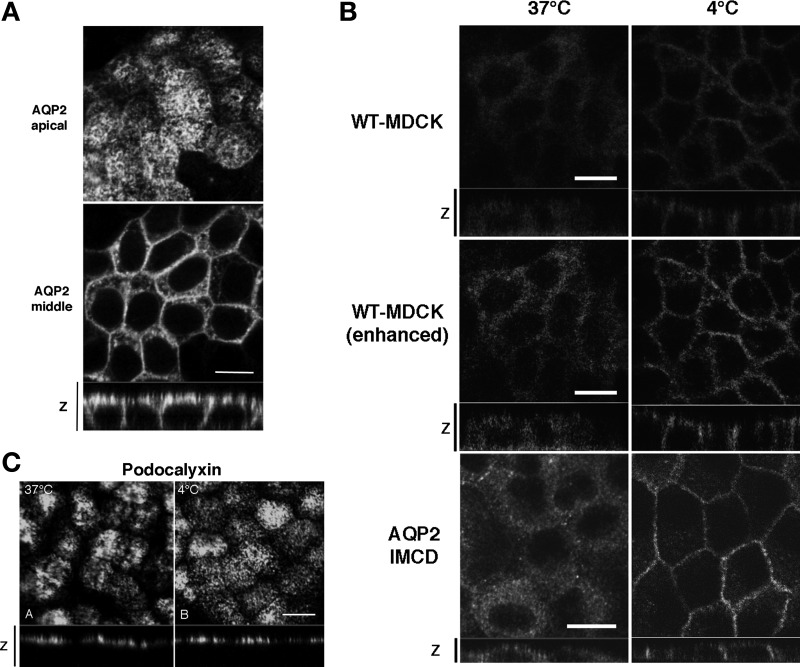
Fig. 2.
The effect of cold shock on subcellular distribution of AQP2. A: AQP2-MDCK cells were incubated at 4°C for 15 min (cold shock) and were then stained for AQP2. The larger panels represent confocal sections through the apical (top) or middle (bottom) regions of the cells as indicated. The smaller horizontal strip at the bottom is a z-section to show apical and basolateral membranes. AQP2 staining was abundant in the basolateral membrane after cold shock. Bar, 10 μm. B: WT-MDCK cells that express low amounts of endogenous AQP2 (top) and AQP2-transfected inner medullary collecting duct (IMCD) cells (bottom) were subjected to cold shock (right), in addition to AQP2-MDCK cells (shown in A). The middle set of images are duplicates of the top two images and were digitally enhanced in Photoshop to show more clearly the staining pattern in the low-expressing MDCK cells. In both cell lines, cold shock induced a basolateral accumulation of AQP2. Bar, 10 μm. C: immunostaining for gp135/podocalyxin under control conditions (37°C) and after cold shock for 15 min (4°C). The apical location of podocalyxin was not modified by cold treatment. Images are representative of three independent experiments. Bar, 10 μm.

