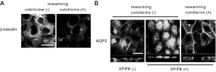
Fig. 7.
Microtubules are required for transcytosis and apical accumulation of AQP2 after VP/FK treatment. AQP2-MDCK cells were treated with or without colchicine (10 μM for 2 h), subjected to cold shock (4°C for 15 min) with or without colchicine, then incubated in DMEM (prewarmed to 37°C) for 15 min at 37°C (rewarming) with or without colchicine. A: after rewarming without colchicine, microtubules were reorganized showing their filamentous structure (left). After rewarming with colchicine, microtubules were maintained in a depolymerized state (right). Bar, 10 μm. B: after rewarming without colchicine, AQP2 was internalized from the basolateral membrane and accumulated in a perinuclear region without VP/FK (left). After rewarming with VP/FK (without colchicine), AQP2 was translocated to the apical membrane with no residual basolateral signal (middle). After rewarming with colchicine and with VP/FK, intracellular AQP2 was increased compared to its location after cold shock alone, but much of the AQP2 still remained in the basolateral membrane (right). The VP/FK-induced apical translocation of AQP2 during rewarming that occurred in the absence of colchicine was not observed in the presence of colchicine (compare right to middle panel). The larger panels represent confocal sections through the middle (left and right) or apical (middle) regions of the cells. Bar, 10 μm. The images are representative of three independent experiments.

