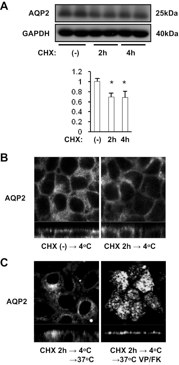
Fig. 8.
The effect of cycloheximide (CHX) on cold shock-induced and rewarming-induced AQP2 subcellular localization. A: AQP2-MDCK cells were treated with 40 μM CHX for 2 h or 4 h, then subjected to Western blotting. Protein band intensity was analyzed by ImageJ. After CHX treatment, AQP2 expression was significantly decreased (30%). AQP2 signal intensity was adjusted to the GAPDH signal and analyzed by a two-tailed Student's t-test (means ± SD, n = 3, *P < 0.05). B: after CHX treatment (40 μM for 2 h), AQP2-MDCK cells were subjected to cold shock (4°C for 15 min). The cold shock induced AQP2 basolateral accumulation (left) was reproduced even after CHX treatment (right). C: AQP2-MDCK cells were treated with CHX (40 μM for 2 h), subjected to cold shock (4°C for 15 min), then rewarmed (37°C for 15 min) with (right) or without VP/FK (VP 20 nM and FK 50 μM for 20 min; left). After rewarming without VP/FK, AQP2 was internalized and accumulated in a perinuclear region (left). After rewarming with VP/FK, AQP2 accumulated in the apical membrane (right). The larger panels represent confocal sections through the middle (top in B and left in C) or apical (right in C) regions of the cells. The images are representative of three independent experiments.

