
Figure 1.
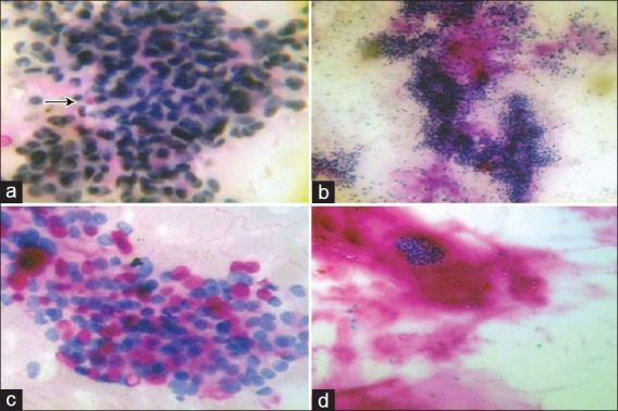
(a) Fibroadenoma showing intracytoplasmic globules (arrow) (PAS-D, ×400), (b) Fibroadenoma showing PAS-D positive clumps of background substance (PAS-D, ×100), (c) Carcinoma breast showing intracytoplasmic globules (PAS-D, ×400), (d) Mucinous carcinoma with abundant background PAS-D positive material (PAS-D, ×100)