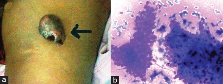
Figure 1.

(a) Focally ulcerated, hyperpigmented polypoid swelling on the dorsal aspect of left thigh. (b) Cellular smear showing epithelial fragments containing small basaloid cells. Fair amount of blue-black pigment is seen in some of the cellular fragments (Giemsa, ×400)
