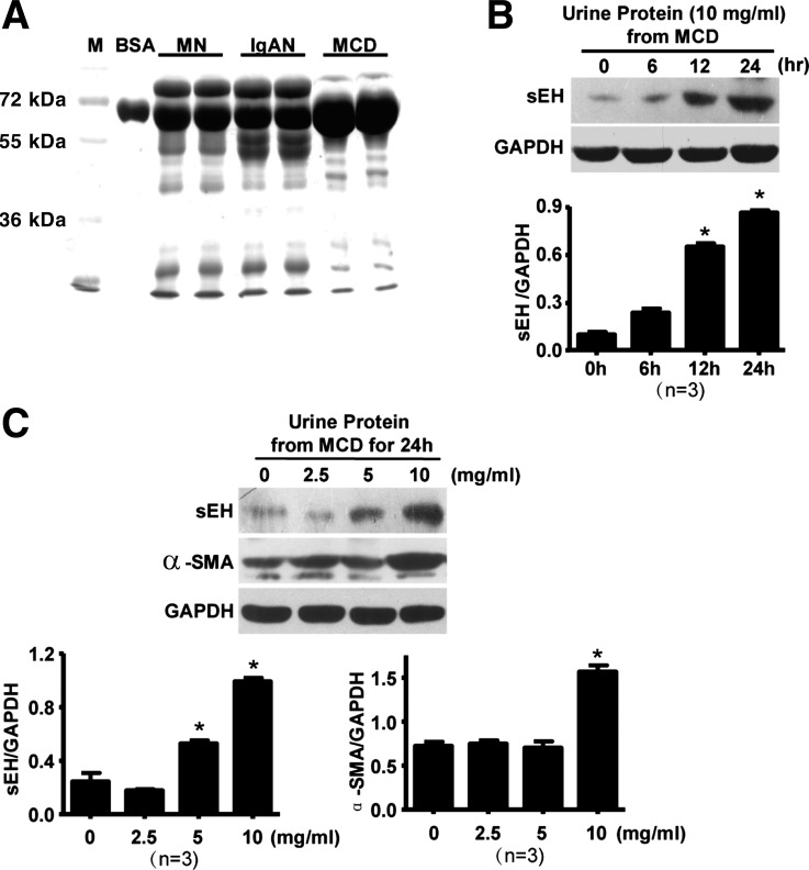
Fig. 2.
Proteinuria in patients with induced sEH expression in cultured rat proximal tubular epithelial cells (RPTECs). A: protein from patients' urine was extracted and analyzed by 10% SDS-PAGE and Coomassie blue staining. Bovine serum album (BSA) was a control. RPTECs were treated with 10 mg/ml proteins from patients with MCD for different times (B) or with different concentrations of urine proteins from patients with MCD for 24 h (C). Western blot analysis of sEH, α-smooth muscle actin (α-SMA), and GAPDH proteins. Expression was normalized to that of GAPDH. Results are representative or means ± SD from at least 3 independent experiments (*P < 0.05 vs. 0 h).

