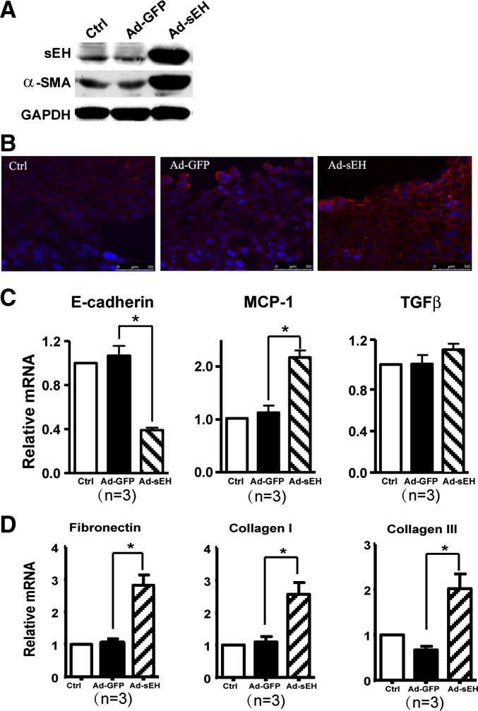
Fig. 4.
Role of sEH overexpression in cultured RPTECs. Confluent RPTECs were infected with recombinant adenovirus encoding sEH (Ad-sEH) or adenovirus encoding green fluorescent protein (Ad-GFP) for 48 h. A: Western blot analysis of sEH, α-SMA and GAPDH proteins. B: confocal microscopy of α-SMA in monolayer cells. Cell nuclei were stained by Hoechst (400×). C: real-time RT-PCR quantification of mRNA levels of E-cadherin, MCP-1, and TGF-β1. D: real-time RT-PCR quantification of mRNA levels of collagen I, collagen III, and fibronectin. Data are means ± SD of the relative mRNA normalized to that of GAPDH from at least 3 independent experiments (*P < 0.05).

