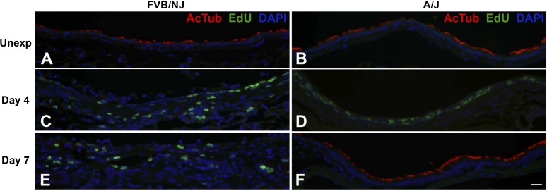
Fig. 11.
Dual fluorescence staining for EdU and ciliated cell marker acetylated tubulin (AcTub). FVB/NJ and A/J mice were exposed to chlorine, and lung sections collected from mice 4 and 7 days after exposure were analyzed by dual staining. Nuclei in proliferating cells are stained green (EdU staining) and AcTub is stained red. Tissues were counterstained with DAPI (blue). Bar in F represents 20 μm for all panels.

