Fig. 3.
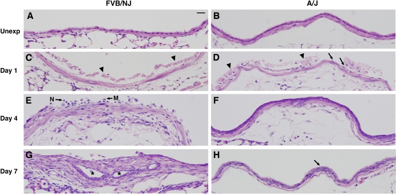
Histological changes in lobar bronchi of chlorine-exposed FVB/NJ and A/J mice. Mice were exposed to chlorine, and lung sections collected from mice 1, 4, and 7 days after exposure were analyzed by hematoxylin and eosin (H&E) staining. A and B are from unexposed mice (Unexp), showing normal structure of bronchi. C and D are from mice at day 1 after exposure, showing massive sloughing of bronchial epithelium in both FVB/NJ and A/J mice (arrowheads) and a thin and flattened layer of epithelial cells remaining in bronchi of A/J mice (arrows in D). E and F are from mice at day 4 after exposure, showing subepithelial thickening with inflammatory cells in the bronchial lumen of FVB/NJ mice (E) and a pluristratified, reparative epithelium in A/J mice (F). N, neutrophil; M, macrophage. G and H are from mice at day 7 after exposure, showing the development of fibroproliferative tissue with epithelial tube formation (asterisks) in FVB/NJ mice (G) and the repair of an injured bronchus by hyperplastic epithelium (arrow) in A/J mice (H). Bar in A represents 20 μm for all panels.

