Fig. 6.
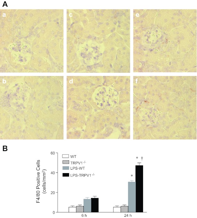
A: representative immunohistochemical staining of F4/80-positive cell in kidney sections from WT and TRPV1−/− mice (a and b) and WT and TRPV1−/− mice treated with LPS (3 mg/kg ip) for 6 (c and d) or 24 h (e and f). Magnification, ×400. B: bar graph shows the number of renal cortical F4/80-positive cell expressed as cells per square millimeter. Values are means ± SE (n = 7 to 8). *P < 0.05 compared with control WT or TRPV1−/− mice; †P < 0.05 compared with LPS-treated WT mice.

