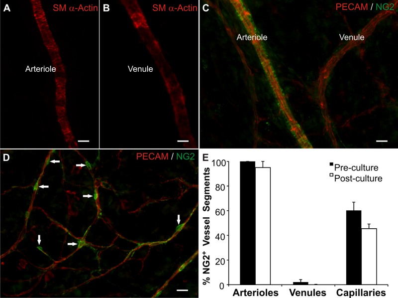
Fig. 1.
Vascular cells remain present and maintain morphological and phenotypic characteristics in the rat mesentery tissues cultured in serum free media for 3 days. After 3 days in culture, arterial (A) and venous smooth muscle cells (B) are identified by smooth muscle α-actin immunolabeling and display respective arterial and venous morphologies. C: neuron-glial antigen 2 (NG2) and platelet endothelial cell adhesion molecule (PECAM) labeling of a paired arteriole and venule. NG2 labeling displayed an arterial-specific smooth muscle cell identity. D: NG2 and PECAM labeling along capillaries. NG2-positive cells exhibited typical pericyte morphology (arrows). E: quantitative comparison of the percentage of vessels with NG2-positive cells in freshly harvested tissues (preculture) and tissues cultured in MEM for 3 days (postculture). Values are means ± SE. Scale bars = 25 μm.

