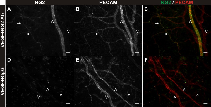
Fig. 6.
Confirmation of antibody targeting NG2-positive cells over the culture time course. A–C: NG2 and PECAM labeling of rat mesenteric tissues post 3 days in culture in MEM + VEGF + NG2 antibody. Anti-rabbit IgG secondary labeling at day 3 identifies NG2-positive smooth muscle cells tightly wrapped around PECAM-positive arterioles and pericytes (arrows) elongated along PECAM-positive capillaries. D–F: NG2 and PECAM labeling in tissues cultured in MEM + VEGF + rabbit IgG antibody. Secondary antibody labeling targeting bound rabbit-derived protein is absent in field of views containing PECAM-positive vessels. “A” indicates arteriole. “V” indicates venule. “c” indicates capillary. Scale bars = 25 μm.

