Fig. 7.
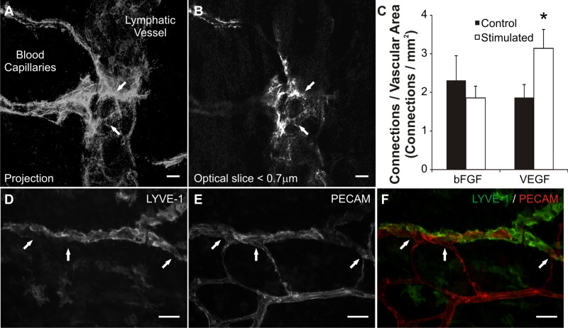
The effect of growth factor stimulation on lymphatic/blood endothelial cell connections. A: confocal projection of sub-0.7-μm slices displays lymphatic/blood endothelial cell connections (arrows) identified by continuous PECAM labeling across cell types. Continuous PECAM labeling was confirmed by observation of each individual optical slice (B) throughout the thickness of the vessels at the connection site. C: quantification of connections per vascular area for bFGF and VEGF stimulated networks. VEGF stimulated an increase in connection density compared with control tissues. The density of connections did not change between unstimulated and bFGF stimulated tissues. *Significance against the control group (P < 0.05). D–F: LYVE-1 and PECAM labeling of lymphatic and blood vessels at connection sites (arrows). Scale bars = 10 μm (A, B), 50 μm (D–F).

