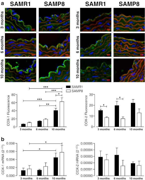
Fig. 4.
Cyclooxygenase (COX) expression in thoracic aorta from SAMR1 and SAMP8 female mice at different ages: atop: representative immunofluorescent merged images of COX-1 (left) and COX-2 (right). Staining shows nucleus (blue, DAPI), actin fibers (red, phalloidin), COX (green). Bar graphs (bottom) show the results of densitometric analyses from pooled data of endothelial expression of COX-1 and COX-2. b COX-1 (left) and COX-2 (right) mRNA expression in mice aorta normalized to the expression of GAPDH, which was used as an endogenous reference gene. Data are plotted as the mean ± SEM derived from four to eight independent experiments. *P < 0.05; **P < 0.01; ***P < 0.001

