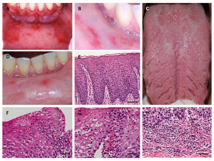
Figure 1.
Clinical and histopathological aspects of geographic stomatitis. (A) Erythematous lesions surrounded by a raised white circinate on the labial mucosa. (B) Partial regression of the lesions was achieved six months after the administration of topics corticosteroids. (C) Fissures on the lingual dorsum. (D) Aspect of lesion after one year. (E) Histopathological features of labial mucosa exhibiting parakeratosis, acanthosis, elongation, and fusion of rete ridges, suprapapillary thinning, and dilated tortuous vessel at the tip of dermal papillae (hematoxylin and eosin (H&E) scale bar in 200µm). (F) Exocytosis of polymorphonuclear leukocytes and Munro microabscesses (H&E, bar in 100 µm). (G) Dilated tortuous vessels and pustule of Kogoj (H&E, bar in 100 µm). (H) Superficial and perivascular lymphocytic chronic inflammatory cell (H&E, bar in 100 µm).

