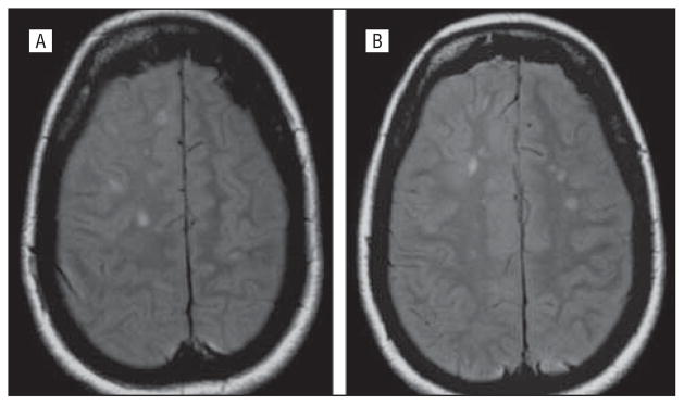
Figure 2.
Magnetic resonance imaging of the brain of patient VI-10, who at age 27 years developed progressive optic atrophy in her right eye without polyneuropathy recognized. She was subsequently diagnosed as having multiple sclerosis when increased T2 signal changes in bilateral white matter paraventricular centrum semiovale regions were found that did not enhance by gadolinium. These changes did not progress, despite new left-eye progressive optic atrophy, characteristic of earlier reported brain imaging in Charcot-Marie-Tooth type 2A2.3,16 Axonal sensory motor polyneuropathy was subsequently diagnosed by clinical and electrophysiologic testing, and genetic testing confirmed her mitofusin 2 Leu146Phe mutation.

