Figure 1.

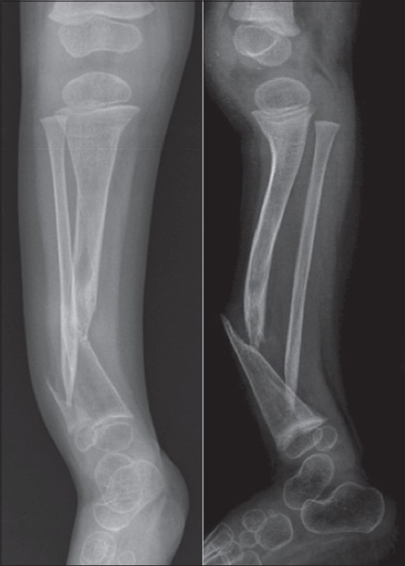
Anteroposterior and lateral radiographs of a patient with CPT. Radiographs show thin and atrophic tibial bone, with a pointed distal fragment. The false joint is in the distal third of the shaft. The fibula is affected

Anteroposterior and lateral radiographs of a patient with CPT. Radiographs show thin and atrophic tibial bone, with a pointed distal fragment. The false joint is in the distal third of the shaft. The fibula is affected