
Figure 1.
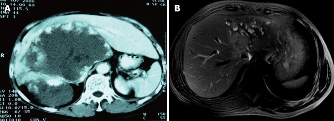
Computed tomography and magnetic resonance imaging of tumor. A: Preoperative computed tomography scan (case 1) demonstrated a large hemangioma located in S1, S4, S5, S6, S7 and S8 of liver, and inferior vena cava (IVC) was involved and compressed severely (IVC circumference 190°, longitude 5 cm); B: Magnetic resonance imaging (case 3) showed the tumor located in S1, S5, S7 and S8, with a diameter of 5 cm; IVC (circumference 60°, longitude 2 cm), right hepatic vein thrombus, main hepatic vein as well as right portal vein were infiltrated by the tumor.