
Figure 1.
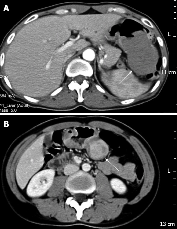
Computed tomography images showed rounded masses in the stomachs, with homogeneous (n = 3) or heterogeneous (n = 1) internal contrast enhancement. A: Contrast-enhanced computed tomography (CT) showed a solitary, exophytic, soft, internal homogeneous tissue mass (arrow) in the greater curvature of the stomach, the mass exhibited central ulceration (Case 3); B: CT during the portal venous phase of contrast enhancement showed a heterogeneous contrast enhanced mass (arrow) in the body of the stomach (Case 1).