Figure 2.

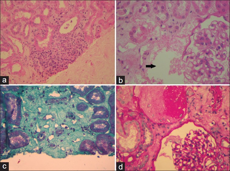
(a) Infiltration of the interstitium by mononuclear cells. (b) Arrow points to an area of tubular necrosis. (c) Interstitial fibrosis appreciated with Massons Trichrome. (d) Glomerulus demonstrating mesangial proliferation

(a) Infiltration of the interstitium by mononuclear cells. (b) Arrow points to an area of tubular necrosis. (c) Interstitial fibrosis appreciated with Massons Trichrome. (d) Glomerulus demonstrating mesangial proliferation