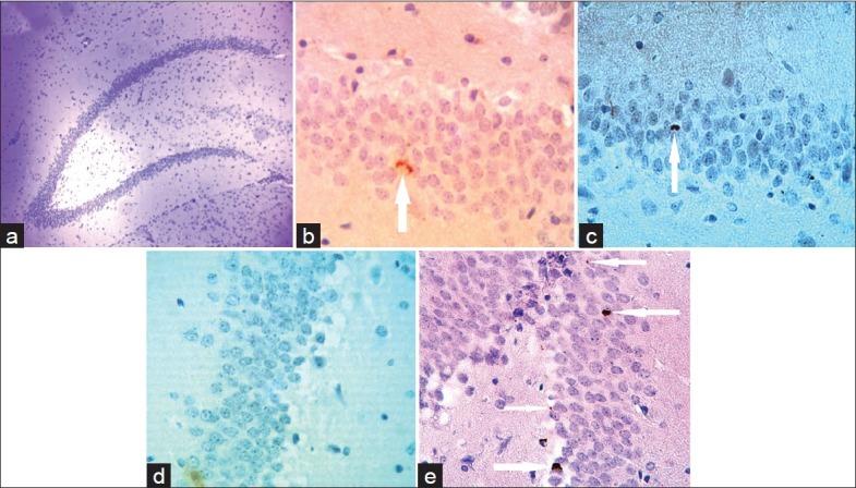
Figure 3.

Photomicrograph of optical microscope from the granular layer of the hippocampal dentate gyrus of rats (a) (M=×4). Ki67-positive cells in the granular layer of the studied groups; control (b), control-erythropoietin (EPO) (c), Alzheimer (d) and Alzheimer-EPO (e) are visible on the arrow tip (M=×40)
