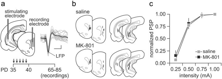
Figure 1.
a, Diagram depicting the recording arrangement used to study the impact of MK-801 exposure on ventral hippocampal-induced synaptic responses in the medial PFC (infralimbic and prelimbic regions) in vivo. Traces are examples of hippocampal stimulation-induced LFP responses recorded in the medial PFC. Calibration: 2 mV, 50 ms. Bottom inset shows the timeline of the experimental design. All electrophysiological recordings were conducted in adulthood (i.e., P65–P85) after noncontingent periadolescent saline or MK-801 (0.1 mg · kg−1 · d−1 for 5 d) injections. b, Diagram summarizing the location of the recording and stimulation sites as determined by histological analyses from Nissl-stained sections. c, Ventral hippocampal-induced LFP responses in the PFC at different current intensities recorded from adult rats with a history of saline (n = 12) or MK-801 (n = 10) treatment during periadolescence. Data are mean ± SEM. PD, Postnatal day.

