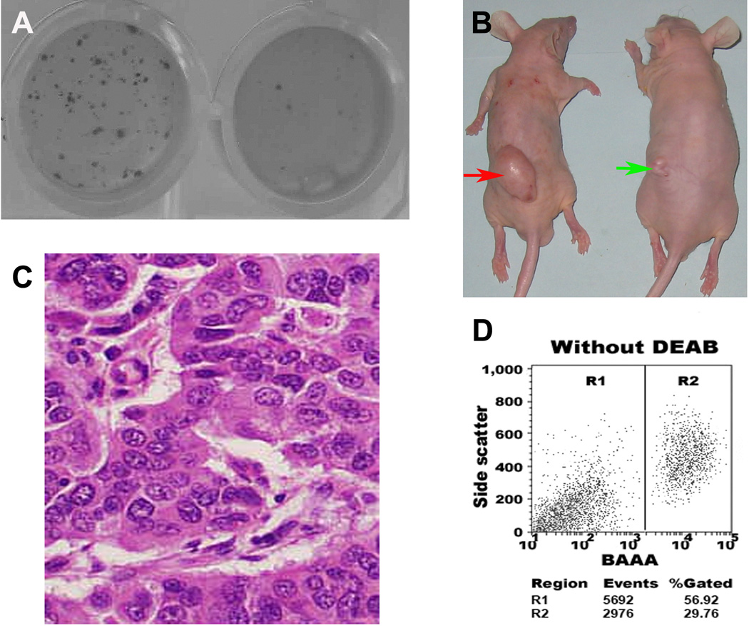
Fig. 2.
ALDH1A1+ bladder cancer cells had high in vitro and in vivo tumorigenic potential. A. ALDH1A1+ cancer cells (left) possessed significantly higher colony-forming efficiency compared with ALDH1A1− populations (right). ALDH1A1+ and ALDH1A1− cells were plated in six-well dishes coated with a thin layer of agar. Three weeks after plating, ALDH1A1+ cells formatted larger and more colonies as compared with the ALDH1A1− cells. B. Tumor formation ability of ALDH1A1+ cancer cells was greater than that of isogenic ALDH1A1− cells. ALDH1A1+ and ALDH1A1− HTB-2 cells were implanted into flanks of nude mice. After four weeks, the dose of 1 × 10⁵ ALDH1A1+ cells yielded larger tumors (red arrow) in all 10 mice with diameters of 32±2.6 mm3, whereas the same dose of ALDH1A1− cells only generated a small tumor mass (5.5 mm3) (green arrow) in only one mouse. The animal experiments were done by using all cell lines. The Fig. 2B only showed the result from HTB-2 cell line. C. Histopathologic examination of the engrafted tumors formed by the ALDH1A1+ HTB-9 cancer cells revealed a highly cellular mass with characteristics of bladder transitional cell carcinoma. D. ALDH1A1+ cancer cells created bladder tumors with heterogeneity in vivo. Reanalyzing cells of the engrafted tumors generated from the ALDH1A1+ HTB-9 by using the Aldefluor assay showed that the xenograft tumors produced 56.9% ALDH1A1+ cells and 29.7% ALDH1A1− cells. The experiments were undertaken on all bladder cancer cell lines and repeated three times. The Fig. 2D only showed the result from HTB-9 cell line.

