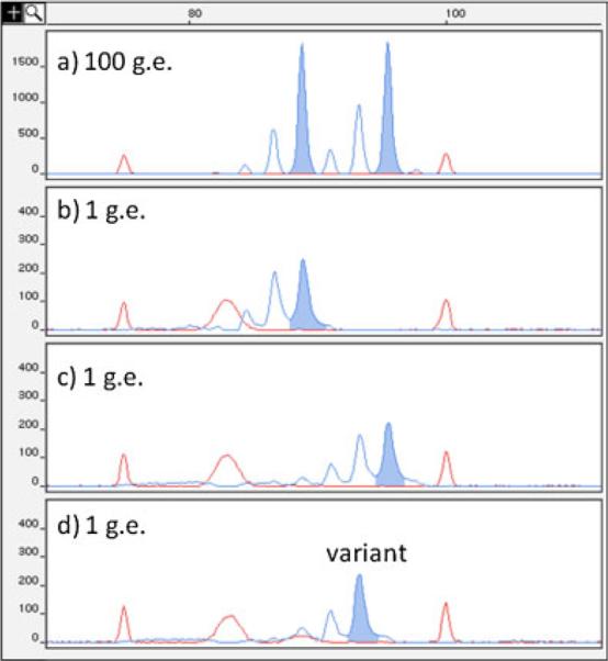
Figure 1.

Representative small pool PCR chromatograms of patient H3 PBL at the D17S518 locus. Patient H3 at D17S518 is an (89, 95) basepair (bp) heterozygote, demonstrated by the presence of both alleles in the 100 genome equivalent genotype in a. Red peaks are internal size standards at 75 and 100 bp. Also seen is a nonspecific electrophoretic band in the size standard mix near 85 bp. After single-molecule PCR, the two progenitors at this locus are seen separated into individual wells in b (89 bp) and c (95 bp). A variant is observed alone in d (93 bp), separated from either progenitor allele.
