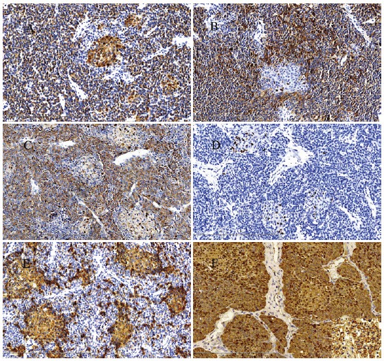
Figure 3.

The tumor cells were positive for Cytokeratin(pan) A, Syn B and CgA C (Original magnification ×200). D: The tumor cells were negative for AFP, but Scattered cells in the squamous corpuscles showed a nuclear staining (Original magnification ×200). E: CD10 immunohistochemistry highlighting the squamoid corpuscles. (Original magnification ×200). F: β-catenin staining exhibited a mixed nuclear and cytoplasmic pattern of the tumor cells. (Original magnification ×200).
