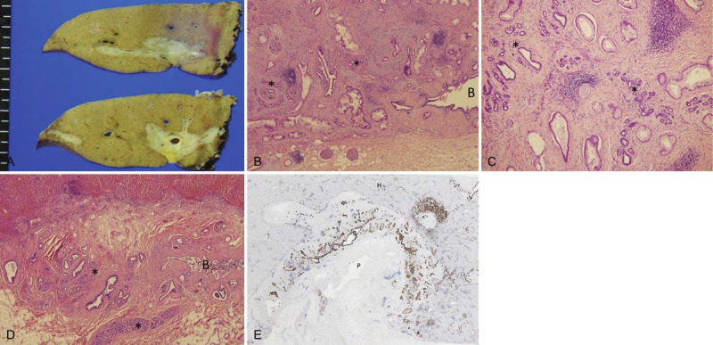
Figure 1.

A: Grossly, the tumor presents as a nodular sclerosing carcinoma of the left hepatic lobe, mainly involving the hilar and perihilar area; B, C, D. Histologically, there were areas of well differentiated adenocarcinoma, mainly extending into pre-existing peribiliary glands. HE. B: Carcinoma mainly involved the pre-existing peribiliary gland network (*). Carcinoma infiltrated the duct wall but not the lumen of perihilar bile duct (B). C: Carcinoma spread into the pre-existing peribiliary glands (*). D: Un-involved peribiliary glands (*) network and bile duct (B) in the same case; E: CK7 immunostaining which was positive in carcinoma cells and also non-neoplastic biliary cells, showed the preferential growth of carcinoma along the peribiliary glands. B, perihilar bile duct; P, portal vein; H, hepatic parenchyma. CK7 immunostaining with hematoxylin counterstaining.
