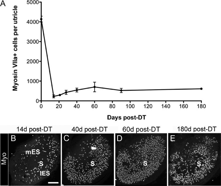
Figure 3.
Many hair cells are replaced after in vivo DT treatment. Hair cell replacement was assessed qualitatively and quantitatively in adult Pou4f3+/DTR mice that received two DT injections. A, Graph shows mean numbers of myosin VIIa+ cells (±SD) per utricle in Pou4f3+/DTR mice at several times post-DT. Sample numbers for counts are provided in Table 1. B–E, Confocal brightest point projection images of myosin VIIa (Myo) labeling in utricles at 14 days (d) (B), 40 days (C), 60 days (D), and 180 days (E) post-DT. S, mES, and lES indicate the presumed position of the striola, medial extrastriolar region, and lateral extrastriolar region, respectively. Scale bar: (in B) B–E, 100 μm.

