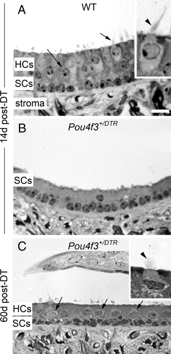
Figure 7.

Hair cell loss and replacement are evident in sectioned utricles. A–C, Cross-sections through the presumed lateral extrastriola of utricles from a wild-type (WT) mouse at 14 days (d) post-DT (A), a Pou4f3+/DTR mouse at 14 days post-DT (B), and a Pou4f3+/DTR mouse at 60 days post-DT (C). The hair cell layer (HCs) and supporting cell layer (SCs) are indicated, as is the stromal layer (stroma). Arrows in A and C point to hair cells. Insets in A and C show exemplary hair cells at higher magnification, with arrowheads indicating the stereociliary bundle in each cell. Scale bar (in A) A–C, 12 μm; A and C insets, 6 μm.
