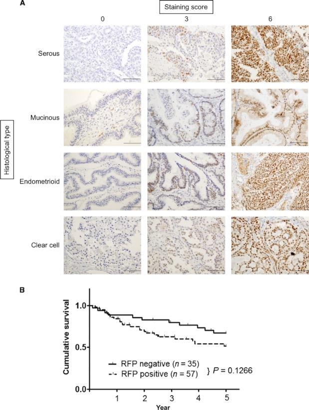
Figure 1.

Immunohistochemical analysis of RFP in epithelial ovarian cancer. (A) RFP expression in serous, mucinous, endometrioid, and clear cell adenocarcinomas. To evaluate RFP expression, the staining intensity was scored as 0 (negative), 1 (weak), 2 (medium), or 3 (strong). The staining extent was scored as 0 (<10%), 1 (10–30%), 2 (30–50%), or 3 (>50%) in relation to the entire cancer area. The sum of scores for the staining intensity and staining extent was used as the staining score (0–6) for RFP. Staining score 0–2, negative; 3–6, positive. Representative images with staining scores 0 (left), 3 (middle), and 6 (right) are shown. Scale bars: 200 μm. (B) Kaplan–Meier survival curves of patients with epithelial ovarian cancer stratified by RFP expression. The 5-year overall survival rate: all cases (n = 92).
