
Fig. 5. PKCδ regulates HIF-1α levels in PC-3 cells via NADPH oxidase.
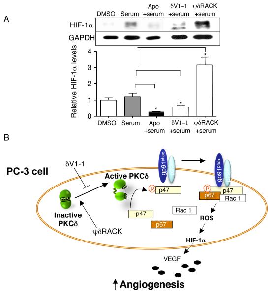
(A) PKCδ regulates HIF-1α levels via NADPH oxidase. PC-3 cells were serum starved for 14 hours and incubated with apocynin (an anti-oxidant and a chemical inhibitor of NADPH oxidase, 1mM) for 5 minutes in the presence of 1% serum (Figure 5A, n=3 each, *; p<0.05). Also, the PC-3 cells were treated with δV1-1 and ψδRACK at 1μM for 4 hours in the presence of 1% serum. The peptides were treated every 1.5 hours and the cells were lysed for Western blot analyses. (B) A schematic diagram summarizes the regulation of tumor-induced angiogenesis via HIF-1α levels by PKCδ and NADPH oxidase in PC-3 cancer cells. p47; p47phox and p67; p67phox.