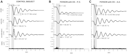
Fig. 2.

Examples of kinematic and EMG traces recorded in one subject without Down syndrome (DS; A) and in two subjects with DS (subjects F.O. and A.A.; B and C). Shadow areas highlight the first flexion. RI, relaxation index; B, damping coefficient; MV, mean velocity during the first flexion; MA, mean acceleration during the first flexion.
