
Fig. 7.
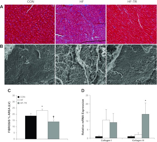
LV fibrosis and relative mRNA expression of collagen I and II isoforms. A: representative histological sections of trichrome-stained LV demonstrating increased fibrosis in HF animals (magnification: ×40). B: representative scanning electron microscopy (SEM) micrographs showing increased quantity and thickness of collagen fibers on the myocardium of the HF group (magnification: ×4,500). C: exercise training prevents increases in LV fibrosis, as indicated by the percent area stained. *P < 0.05, HF vs. Con and HF-TR. †P < 0.05, HF-TR vs. Con. D: collagen III mRNA expression is increased in the LV of HF-TR compared with Con and HF animals. *P < 0.05, HF vs. HF-TR and Con. Values are means ± SE.