Figure 1. .

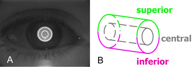
OCT scan pattern for imaging the anterior chamber of the eye. (A) Concentric circular scans of 2- and 4-mm diameters were overlaid on a charge-coupled device image. (B) Data from the central, superior, and inferior regions were analyzed.

OCT scan pattern for imaging the anterior chamber of the eye. (A) Concentric circular scans of 2- and 4-mm diameters were overlaid on a charge-coupled device image. (B) Data from the central, superior, and inferior regions were analyzed.