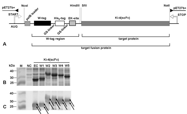
Figure 2.

Cloning scheme for Wx-Ki-4(scFv) fusion protein constructs, SDS-PAGE and Western blot analysis of Wx-Ki-4(scFv) expression. (A) Plasmid map of pET-Wx-Ki-4(scFv). The plasmid backbone contains a kanamycin resistance gene (kan), a pBR322 origin of replication and the lactose repressor gene (lacI). The Ki-4(scFv) sequence is genetically linked to the W-tag. The schematic structure of the Wx-Ki-4(scFv) insert in the expression cassette consists of the pelB signal peptide inducible with IPTG via the lac operator. The W-tag is fused to a His6-tag by a GS-linker and the Ki-4(scFv) is linked to the His6-tag through a cleavable enterokinase site. Restriction sites are also shown. (B) SDS-PAGE: lane 1 – Prestained Broad Range Protein Marker (NEB, USA), lane 2 – negative control (NC) (pET-27b+ vector), lane 3 – expression control (EC) (Ki-4(scFv)), lane 4 – W1-Ki-4(scFv), lane 5 – W2-Ki-4(scFv), lane 6 – W3-Ki-4(scFv), lane 7 – W4-Ki-4(scFv), lane 8 – W5-Ki-4(scFv). (C) Western blot of proteins detected with an anti-poly-His antibody, a goat anti-mouse IgG peroxidise conjugated antibody and DAB. In figures (B) and (C) the proteins are detected as double bands. The lower band (25-30 kDa) corresponds to the calculated weight of the protein, the higher band (30-35 kDa) includes the uncleaved pelB leader peptide.
