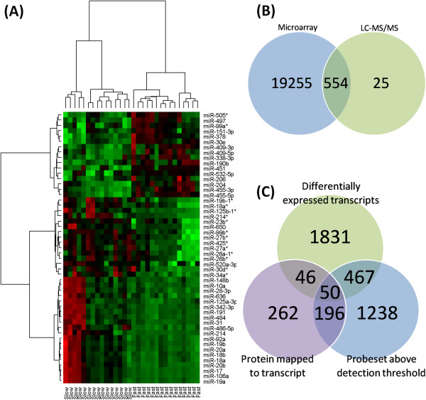
Figure 2.

miRNA DE analysis heatmap and mRNA-proteomic mapping. miRNA expression profiling identified 51 miRNAs that were DE and correlated to sample growth rate. (A) HCA analysis confirmed that those DE and correlated genes separate the samples into fast and slow groups (red indicates diminished miRNA expression and green indicates increased miRNA expression). (B) 260 DE proteins mapped to one or more probesets. (C) 196 probesets corresponding to 158 DE proteins expressed above the detection threshold and unchanged at the mRNA level between the fast and slow groups.
