
Figure 2. Transplantation of melanoma cells into three distinct niches of the chick embryo.
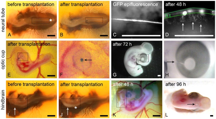
(A) Chick embryo stage 12–13 HH before and (B) directly after transplantation of B16-F1 melanoma cells into the neural tube. The entering site of the micro-pipette is marked (asterisk in A). Note the dilated neural tube (frame in B) due to the transplanted cells when compared to (A). (C) B16-F1 cells can be detected via GFP epifluorescence in the lumen of the neural tube directly after transplantation. (D) 48 h after transplantation ventrally emigrating B16-F1 cells are clearly discernible (arrows) in lateral view; the borders of the neural tube are outlined in green. (E) Chick embryo stage 19 HH directly after transplantation of B16-F1 melanoma cell aggregates into the optic cup. (F) Aggregates were stained with nile blue sulphate before transplantation for better visibility. Higher magnification shows the aggregates behind the embryonic lens (arrow). A temporary capillary bleeding can be discerned at the injection spot at the choroid fissure in (F). (G) Macroscopically no tumor growth is visible 72 h after transplantation. (H) The former entering site of the micro-pipette (choroid fissure) is marked (arrow). (I) Chick embryo stage 12–13 HH before and (J) directly after transplantation of human melanoma cells into the ventricle of the hindbrain (rhombencephalon, frame in I). The entering site of the micro-pipette is marked with the asterisk in (J). Note the melanoma cell-filled brain ventricle (frame in J). (K) 48 h after transplantation a growing tumor is already visible in the hindbrain (frame). (L) After 96 h a single condensed tumor is visible in the dorsal midline of the neural epithelium (arrow). Scale bars in A, B and E–L: 1 mm; scale bars in C and D: 0.5 mm.