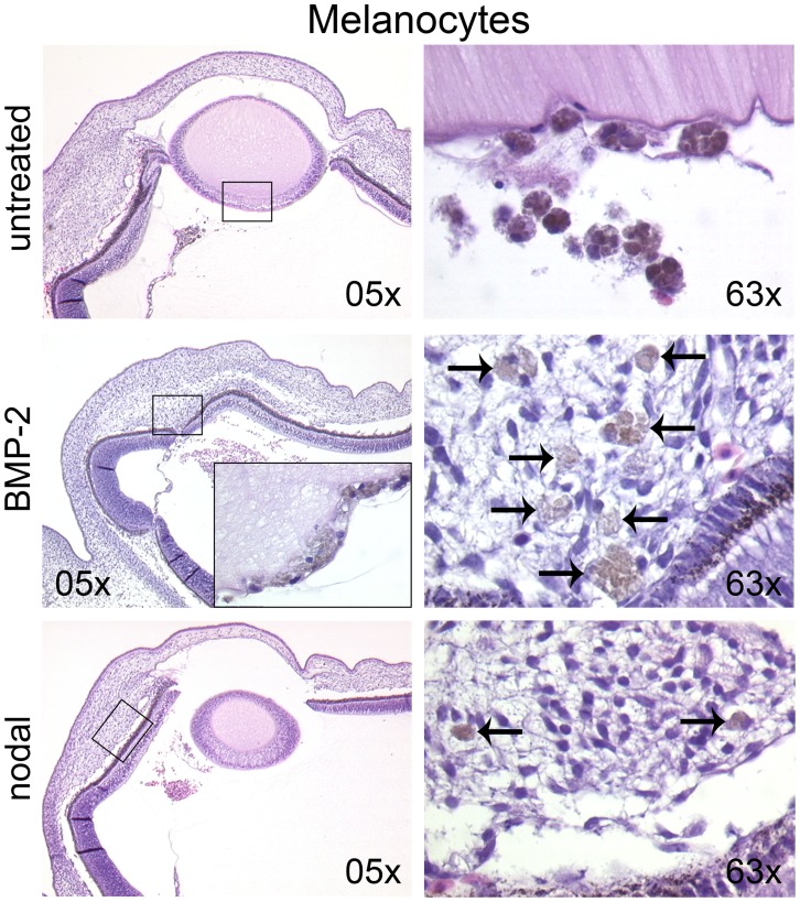
Figure 4. Pre-treatment with the TGFbeta family members BMP-2 or nodal induces invasive migration of human melanocytes in the optic cup.

Untreated, BMP-2 or nodal pre-treated melanocytes were injected into the optic cup of the chick embryo (stage 20 HH). After 72 h of further incubation, the embryos were analyzed for tumor growth and invasion. Untreated melanocytes formed loosely aggregated tumors adjacent to the hyaloid vessels (left image in upper row), in the developing vitreous body and behind the lens (right image in upper row) without invasion. The BMP-2 and nodal groups formed tumors in similar locations. In the BMP-2 group single melanocytes invaded the lens epithelium (insert in left image in middle row), the retina, the hyaloid vessels, and the choroid (right image in middle row; arrows pointing at melanocytes). In the nodal group single melanocytes invaded the choroid (lower row, arrows in right image) and the hyaloid vessels.
