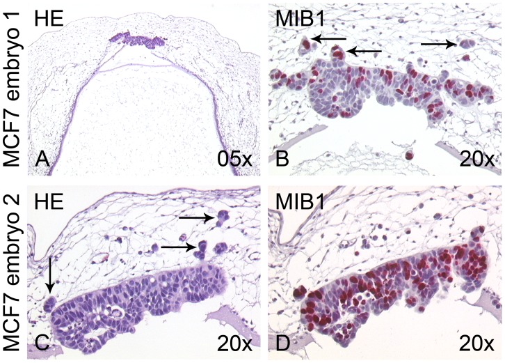
Figure 5. Transplantation of MCF7 breast cancer cells into the rhombencephalon of the chick embryo.

(A,C) Two different examples of chick embryos 96 h after transplantation of MCF7 breast cancer cells into the rhombencephalon, H&E stainings. The cells have formed stretched, compact epithelial tumors in the roof plate and adjacent mesenchyme. (B,C) Higher magnification reveals that the MCF7 cells invade the chick host in small aggregated clusters (arrows). (B,D) MIB1 immunohistochemistry shows that 30–50% of the MCF7 cells proliferate in the chicken environment.
