
Figure 3. Forebrain and hindbrain pax2a:GFP positive cells are sensitive to ethanol exposure.
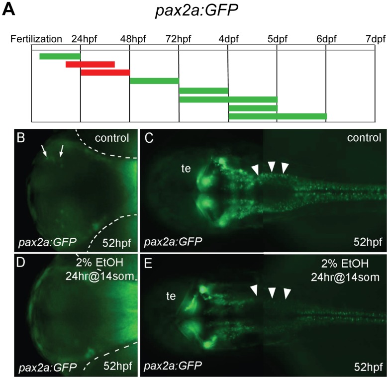
A, the duration and embryonic stage of 2% ethanol treatments illustrated with colored bars for Tg(pax2a:GFP) embryos, as described in Fig. 1. B−E, ventral views (B, D) and dorsal views (C, E) of Tg(pax2a:GFP) embryos at 52 hpf. B, C, are controls. D, E, were treated with 2% ethanol for 24 hrs at the 14 somite stage. B, D, a small, distinct group of neurons in the forebrain (arrows in B) were missing after ethanol exposure (D). The location of the eyes is outlined (dashed white lines). C, E, neurons around the midbrain-hindbrain region were mostly unaffected, hindbrain neurons were drastically reduced (arrowheads), and spinal cord neurons appeared slightly reduced. Tectum (te).