
Figure 6. Expression of GFP in Tg(oxtl:GFP) embryos corresponds exactly with the expression of endogenous oxtl mRNA.
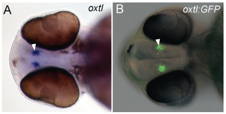
A,B, ventral view of 48 hpf embryos. A, whole-mount in situ hybridization for oxtl mRNA in a 48 hpf embryo. B, GFP expression visualized with epi-fluorescence in a 48 hpf Tg(oxtl:GFP) embryo. Epi-fluorescent and differential interference contrast (DIC) images are overlaid. The oxtl expressing cells are indicated with arrowheads.