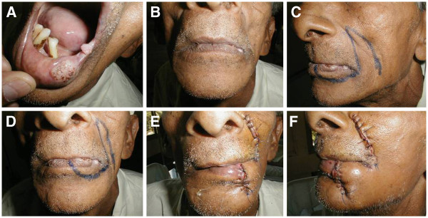
Figure 1.

Clinical photographs showing surgical procedure for inserting nasolabial flap. (A) Two discrete lesions on the lower lip and commissure. (B) Front view of patient with mouth closed. (C) Lateral profile, showing incision. (D) Front view of the incision. (E) Front view after completion of surgery and insertion of flap. (F) Lateral profile after completion of surgery and insertion of flap.
