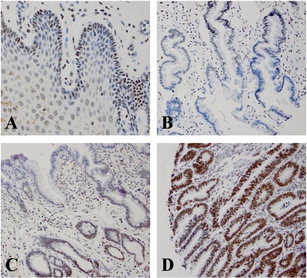
Figure 1.

High Bmi-1 expression in various histologic types by immunohistochemical studies. A. Bmi-1 positive cells predominantly in the basal layer of normal esophageal squamous epithelium; B. Distribution of Bmi-1 positive cells mostly at the base of glands in columnar cell metaplasia; C. Bmi-1 positive cells mostly at the base of glands in intestinal metaplasia; D. Distribution of Bmi-1 positive cells evenly in the glands of low-grade dysplasia glands.
